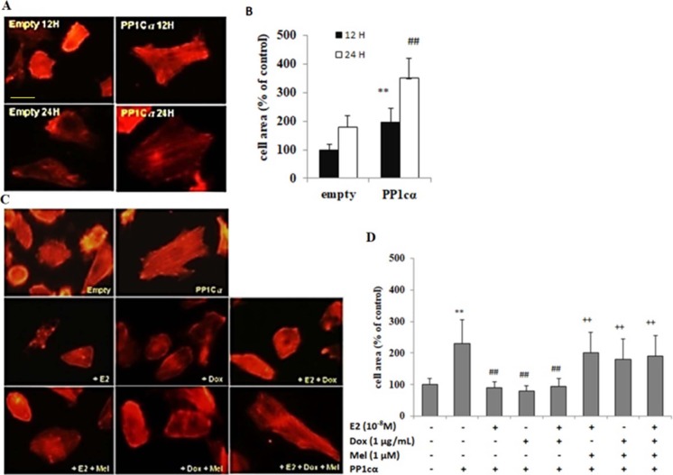
Fig 4. PP1cα overexpression mediated cardiomyoblast hypertrophy was attenuated by E2 and/ or Dox treatment, which was reversed by treatment with specific ERα inhibitor.
A & B, Tet-on/ERα H9c2 cells were transfected with overexpressing vectors carrying PP1cα. At 12 h, actin staining was performed which showed that cell surface area was increased by 2 folds in PP1cα transfected cells as compared to cells transfected with empty vectors alone. At 24 h, further increase in cell surface area was observed by approximately 1.5 folds as compared to 12 h and cell surface area quantification was performed. Student’s t-test, “**”, p < 0.01 for control versus PP1cα 12 h; “##”, p < 0.01 for control versus PP1cα 24 h. C & D, Tet-on/ERα H9c2 cells were pre-treated with either E2 or Dox, or melatonin or combination treatments, followed by transfection with PP1cα overexpressing vectors for 24 h. As evident by actin staining, E2 or Dox or combination treatments reduced hyper growth effect induced by PP1cα overexpression. However, treatment with melatonin reversed the anti-hypertrophic effect of E2, Dox and/ or combination treatments in PP1cα transfected cells. The bar graph (mean ± S.D.) represents data from three independent experiments. 30 cells were counted per condition in each experiment, and the data was analysed by Student’s t-test. “**”, p < 0.05 for untreated versus PP1cα; “##”, p < 0.05 versus PP1cα or “++”, p < 0.01 versus untreated.

