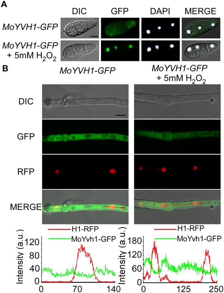
Fig 1. MoYvh1 translocates into the nucleus in response to the oxidative stress.
(A) Fluorescence observation of conidia untreated (upper panels) and treated with 5 mM H2O2 for 2 h (lower panels). 4’,6-Diamidino-2-phenylindole (DAPI) was added to the cultures 5 min prior to the observation of the nuclei. The merged images of GFP and DAPI staining showed that MoYvh1-GFP is localized in the nucleus when treated with H2O2. Bar = 5 μm. (B) Fluorescence observation of mycelia contain MoYvh1-GFP and H1-RFP were untreated (left panels) and treated with 5 mM H2O2 for 2 h (right panels). “green line” represents MoYvh1-GFP, “red line” represents H1-RFP. Insets highlight areas analyzed by line-scan. Bar = 5 μm.

