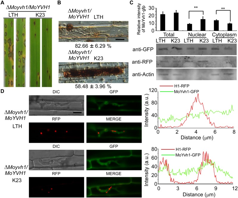
Fig 6. Host derived ROS induces MoYvh1 nuclear accumulation during infection.
(A) Whole-plant assays with cultivars LTH and K23 inoculated with the ΔMoyvh1/MoYVH1 strain. The strain formed rare, small lesions on K23 which were different from the lesions on LTH. Plants were photographed at 7 d after inoculation. (B) DAB staining of the excised leaf sheath of cultivars LTH and K23 infected by the ΔMoyvh1/MoYVH1 strain 30 h after inoculation. Bar = 5 μm. (C) Rice leaves were incubated with ΔMoyvh1/MoYVH1-GFP-H1-RFP strain for 30 h. Equal weight of rice leaves (LTH and K23) were divided into three parts for extraction of total, nuclear and cytoplasm proteins. Equal amounts of total, nuclear and cytoplasm proteins were separated by SDS-PAGE, and the presence of MoYvh1 was detected by Western blotting using the anti-GFP antibody. The intensity of Western blotting bands was quantified with the ODYSSEY infrared imaging system (application software Version 2.1). The intensity of MoYvh1 was compared between the cv. LTH and cv. K23 among total proteins, nuclear proteins, and cytoplasmic proteins. H1 (a nucleus marker) and actin (a cytoplasm marker) were detected by Western blotting analysis using the anti-RFP or anti-Actin antibodies. Bars denote standard errors from three independent experiments. Asterisk indicates significant differences (Duncan’s new multiple range test p < 0.01) (D) Localization of MoYvh1 during infection. Infection hyphae contain MoYvh1 and H1-RFP were observed by confocal fluorescence microscopy in the sheath of cultivars of LTH and K23 at 30 hpi. “green line” represents MoYvh1-GFP, “red line” represents H1-RFP. Insets highlight areas analyzed by line-scan. Bars = 5μm.

