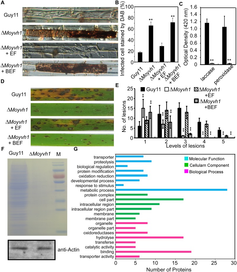
Fig 7. MoYvh1 nuclear localization promotes extracellular proteins to evade host innate immunity.
(A) Extracellular fluids from Guy11 appressoria suppress the defects in scavenging host-derived ROS of the ΔMoyvh1 mutant, whereas boiled EF did not exhibit a similar defense response. “EF” represents the extracellular fluid. “BEF” represents the boiled extracellular fluid. Data represents observations from three independent experiments. Bar = 5 μm. (B) The infected cell stained by DAB. Three independent biological experiments were performed, with three replicates each time and yielded similar results in each independent biological experiment. Error bars represent standard deviation, and asterisks represent significant difference between the different strains (p < 0.01). (C) Laccase activity measured by ABTS oxidizing test without H2O2 and peroxidase activity measured by ABTS oxidizing test with H2O2. Bars denote standard errors from three independent experiments. Asterisk indicates significant differences (Duncan’s new multiple range test p < 0.01) (D) The conidial suspensions of each treatment were sprayed on the rice leaves. “EF” represents the extracellular fluid. “BEF” represents the boiled extracellular fluid. (E) Quantification of lesion type. Lesions were photographed and measured at 7 days post-inoculation (dpi), counted within an area of 4 cm2 and experiments were repeated three times with similar results. Asterisk indicates significant differences at p = 0.01. (F) 1D gel analysis of Guy11 and the ΔMoyvh1 mutant extracellular fluid proteins: Equal numbers of Guy11 and ΔMoyvh1 conidia were harvested and equalized by using the actin antibody. The same number of conidia was used to extract the extracellular fluid. The extracellular fluid was concentrated to 100 μl. A 50 μl aliquot of each sample was then fractionated by 1D SDS-PAGE and proteins were stained by Coomassie brilliant blue. (G) GO functional analysis of extracellular fluid proteins.

