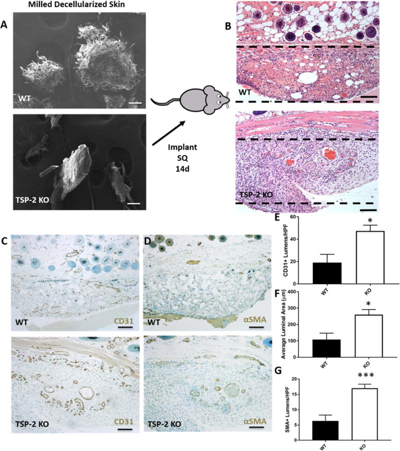
Fig. 4. Comminuted TSP-2 KO ADM promotes vascularization.

SEM of WT and TSP-2 KO powdered ECM indicated difference in the structure of the grains, with WT appearing more shredded (A). After 14 days in vivo the powder was well invaded (B). Immunohistochemistry demonstrated more vascularization around the TSP-2 KO powder as demonstrated by CD31 (C) and αSMA (D). There were more CD31-positive lumens (E) that were larger (F) and more αSMA-positive (G). Scale bars = 100 μm. Implanted silicone trays are out of frame but reside below and to the sides of the implanted ECM. ECM is just below the dermis. Results are given as mean + SEM, n=6, *p<0.05, ***p<0.005.
