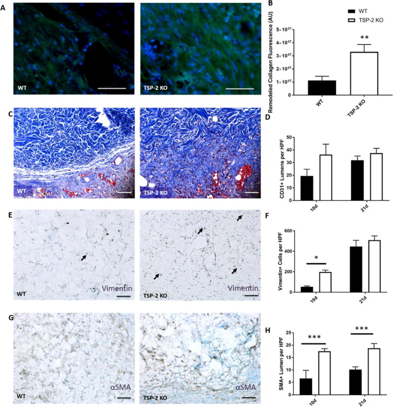
Fig. 6. TSP-2 KO ADM exhibits enhanced integration and vascular maturation in diabetic wounds.

Representative images of collagen remodeling in ADM after 10 days of implantation in diabetic wounds (A) demonstrate increased remodeling in TSP-2 KO ADM (B). Representative images of Masson’s trichrome staining along border of graft demonstrate increased tissue integration with TSP-2 KO ADM; the border between normal tissue and graft is no longer visible by 10 days (C). There are no differences in total vessel number (CD31) between WT and TSP-2 KO ADM treated wounds at 10 or 21 days (D). Representative images of vimentin staining after 10 days (E). Quantification of vimentin stain indicates an increased penetration of mesenchymal cells within the TSP-2 KO ADM by 10 days, but the WT ADM were no different by 21 days (F). Representative images of αSMA after 10 days (G) and quantification revealing more positive vessels at both 10 and 21 days. n=4 (10 days) or n=6 (21 days). Scale bars = 50 μm. Results are given as mean + SEM, *p<0.05, **p<0.01.
