. 2018 May 3;7:e32911. doi: 10.7554/eLife.32911

Figure 6. Bqt4-rich domains ensure faithful inheritance of heterochromatin.

(A) Schematic of system designed to move replication forks to the NE. Coexpression of Polα-GFP and Man1-GBP recruits Polα-associated (replicating) chromatin to Man1-rich subdomains of the NE. Images below schematic show remobilization of Pol α-GFP to colocalize with Man1-GBP-mCherry encircling the nucleus. (B) In cells lacking Man1-GBP, Polα-GFP shows diffuse localization within the nuclear interior. (C) ChIP-seq experiments were performed as described in text and Methods, using H3K9Me2 antibody (ab1220, Abcam). (D) Ribbon line-plot of enrichment of H3K9Me over the ade6+ locus in three independent WT isolates. The solid blue line indicates median; width of the ribbon indicates range of enrichment levels among the three isolates. (E–F) Ribbon line-plot of enrichment of H3K9Me2 at centromere of Chr III. The central core region is indicated by the yellow box. The sharp peaks flanking the core region (F) align to small repetitive regions (~25 bp) present in multiple regions across the genome; it is therefore not possible to determine their source.

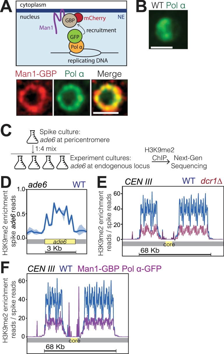
Figure 6.

Figure 6—figure supplement 1. A C-terminal GFP tag on Polα does not interfere with centromeric silencing.

Figure 6—figure supplement 1.

Five-fold serial dilutions of the indicated strains are shown. While wt cells expressing ura4+ grow on media lacking uracil and fail to grow in the presence of FOA, ura4-D18 cells fail to grow on -URA but grow on FOA. Cells harboring ura4+ at the centromeric imr1R locus grow on FOA due to silencing at imr1R; these cells also show a partial loss of viability on -URA due also to imr1R silencing. This silencing is intact in cells carrying Polα/Swi7-GFP.
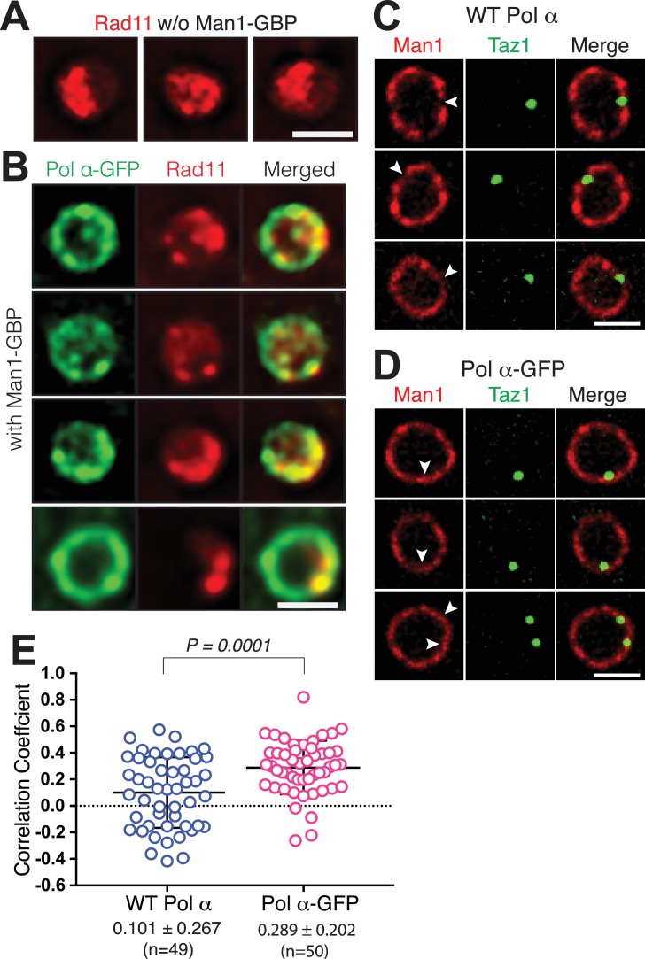
Figure 6—figure supplement 2. Man1-GBP pulls both Polα-GFP and replicating telomeres to Man1-rich domains.

Figure 6—figure supplement 2.

(A) Three representative wt cells showing Rad11-mCherry visible throughout the nucleus (single Z-plane images of live cells). (B) Each row shows a representative live cell expressing Pol α-GFP Man1-GBP and Rad11-mCherry (single Z-plane images of the nuclei). Pol α-GFP remobilizes to the NE, forming a fluorescent ring. Rad11 also appears to remobilize to the NE, with cells having lower detectable Rad11-mCherry signal in the nuclear interior compared to wt cells (compare with A). (C) Taz1 tends to localize to Man1-poor regions (quantified in (E)) in a wt setting. Imaging performed on DeltaVision microscope. (D–E) In cells harboring the Man1-GBP/Polα-GFP tethering system, the frequency of Taz1/Man1 overlap increases. (D) Imaging as in (C). (E) Pearson correlations coefficients defining degree of colocalization between Taz1-mTurquoise and Man1-GBP-mCherry were determined using Applied Precision softWorX software. While Taz1 rarely colocalizes with Man1 in a wt scenario, GBP- mediated recruitment of Polα-GFP pulls replicating telomeres to Man1 sites.