
Figure 1.
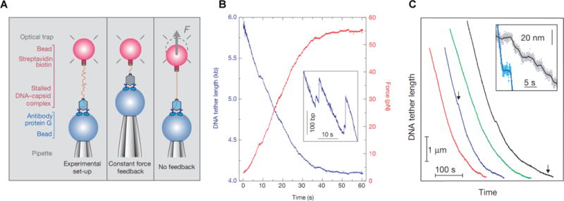
Single-molecule viral DNA packaging assay. A) Schematic of the experimental setup used in the earliest work. A single DNA molecule hanging out of a stalled φ29 packaging complex was tethered at one end to a microsphere held in an optical trap while the procapsid was bound to a second microsphere held by a micropipette. After initiating packaging with ATP two measurement modes were used: “Constant force feedback”, where the separation between the microspheres was adjusted to keep the DNA stretching force constant, or “No feedback” where the separation was fixed and the DNA stretching force was allowed to rise as packaging proceeded. B) Force vs. time (red line) for a packaging event measured without feedback, reaching ~55 pN before the motor paused or stalled, and corresponding tether length vs. time (blue line). Inset is a zoomed view illustrating occasional slipping events where the DNA moved backwards out of the capsid. C) DNA tether length (i.e., unpackaged DNA length) vs. time during packaging with 5 pN force feedback (the four different colored lines indicate four different single packaging events, shifted arbitrarily along the time axis for clarity). D) Inset is a zoomed view of the regions marked with arrows, illustrating occasional pauses in translocation.