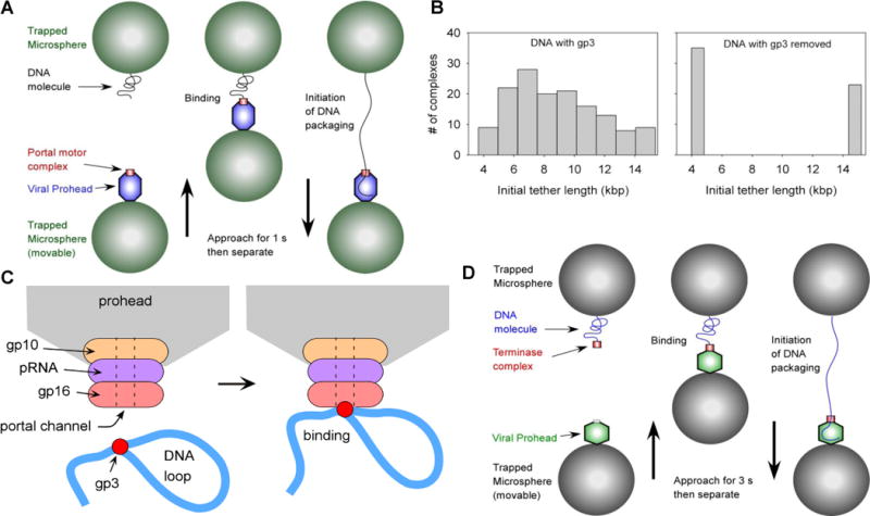
Figure 2.

Measurements of the initiation of viral DNA packaging A) Schematic of the experimental setup used to initiate φ29 packaging in real time. Instead of using a stalled, partially packaged complex as in Fig. 1, a pre-assembled prohead-motor complex is “fed” DNA. Each microsphere was held in a separate optical trap. The same approach was used for initiating phage T4 packaging. B) Measured distribution of initial tether lengths for packaging native φ29 DNA with its gp3 terminal protein, or with gp3 removed by digestion with proteinase K. C) Proposed model for gp3-mediated DNA looping at the initiation of φ29 packaging. D) Modified approach used to initiate single-molecule phage λ packaging. A motor-DNA complex is preassembled and brought into proximity of a λ procapsid (instead of preassembling a motor-procapsid complex).
