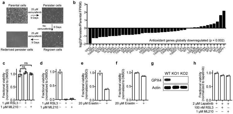
Extended Data Figure 1. Additional data demonstrating persister cell ferroptosis sensitivity.
a, A375 melanoma persister cells are reversibly drug-resistant. Scale bars indicate 400 μm. b, Global antioxidant gene-expression is downregulated in BT474 persister cells. P value calculated using a two-tailed Wilcoxon signed rank test. c, MCF10A parental cells and d, BT474 persister cells derived from carboplatin and paclitaxel were treated with RSL3 or ML210 for three days. e, BT474 persister cells or f, parental cells were treated with erastin for five days. g, Western blot demonstrating GPX4 knockout in two distinct A375 clones (clone 1, KO1; clone 2, KO2). For gel source data, see Supplementary Figure 1. h, BT474 parental cells co-treated with 2 μM lapatinib and RSL3 or ML210 for three days. Data are plotted as means and error bars represent standard deviation. c, n = 3 and d-h, n = 2 biologically independent samples. c, P value calculated from a two-tailed t test where ns represents P > 0.05. All data are representative of two separate experiments.

