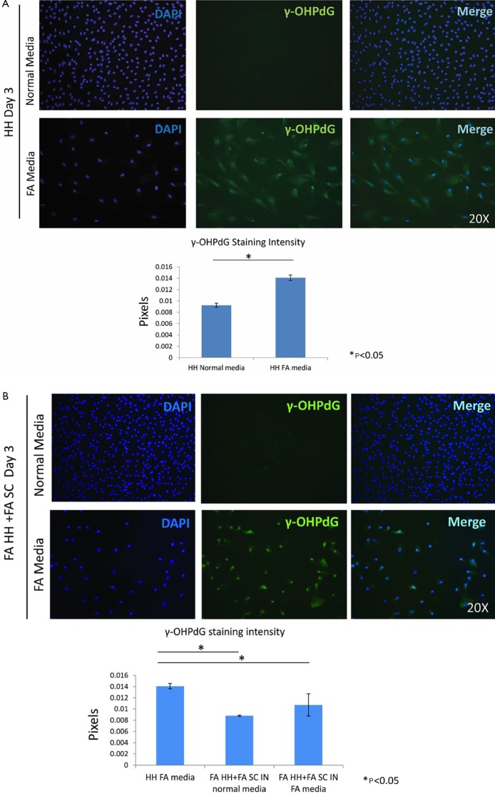
Figure S2.
Co-culture in normal and FA conditions. (A) ICC and quantification of the ICC staining of γ-OHPdG of HH in normal media and FA media after 3 days; (B) fatty acid treated HH and SC in normal media by day 3 and FA-treated HH and FA SC in FA media by day 3 compared to HH in FA media with intensity quantification of γ-OHPdG. * indicates significance (P≤0.05) between groups using two-sample independent t-tests. ICC, immunocytochemistry; FA, fatty acids; HH, human hepatocytes.

