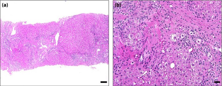
Figure 2.

Liver biopsy specimen showed burned‐out non‐alcoholic steatohepatitis. (a) Distortion of hepatic lobular architecture was extensively observed (hematoxylin–eosin stain; lower‐power view). (a,b) Although few lipid droplets (<5%) were observed, burned‐out non‐alcoholic steatohepatitis10 was considered, because pericellular and perivenular fibrosis were prominent, and other causes of liver diseases were ruled out in the present case. Scale bars represent (a) 100 μm and (b) 20 μm.
