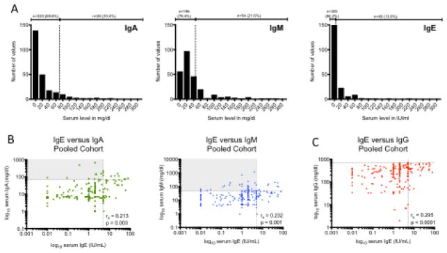
Fig. 2.
IgE compared to other serum Ig isotypes in patients with CVID. A) Distribution of serum IgA, IgM and IgE in CVID. Vertical dotted line represents LLN. B) Correlation of serum IgE with serum IgA and IgM. C) Correlation of serum IgE with serum IgG. Horizontal dotted lines represent LLN; vertical dotted lines represent IgE cutoff of 5 IU/ml. Rs = Spearman r

