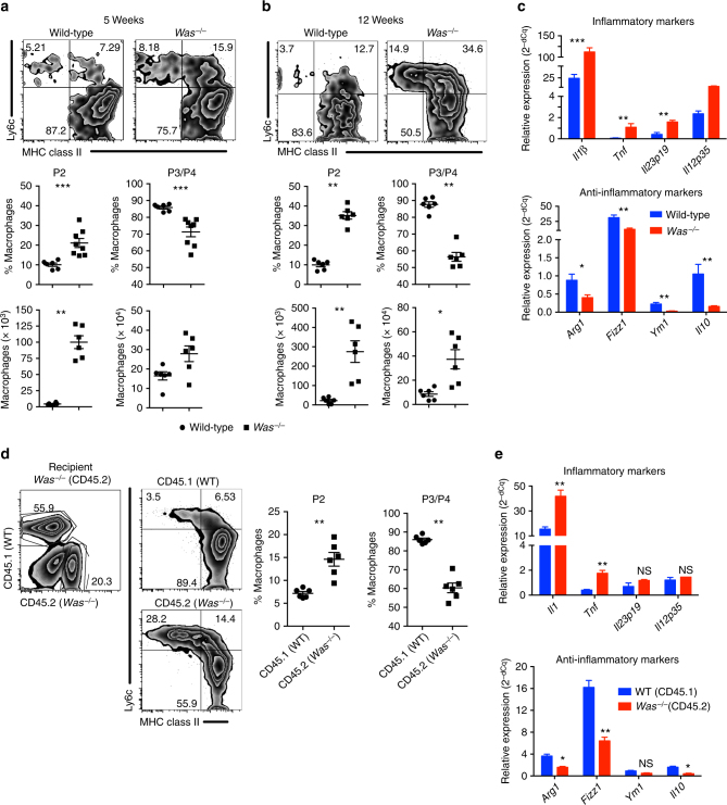
Fig. 1.
Defective anti-inflammatory macrophage differentiation and function in the colon of Was−/− mice. Flow cytometric analysis of LP macrophage in mice at a 5 (WT n = 6; Was−/− n = 8) and b 12 (WT n = 6; Was−/− n = 6) weeks of age followed by quantification of pro- (P2) and anti-inflammatory (P3 + P4) subsets. Macrophages were gated as live CD45+CD11b+CD103-CD64+ cells. Data are cumulative of three independent experiments. *p < 0.05, **p < 0.01, ***p < 0.001 (Student’s t-test). c Expression of pro- and anti-inflammatory genes in sorted P3 + P4 macrophages (WT n = 12; Was−/− n = 12). P3 + P4 cells from three mice were pooled together. Data are cumulative of two independent experiments. *p < 0.05, **p < 0.01, ***p < 0.001 (Student’s t-test). d CD45.1+ (WT) and CD45.2+ (Was−/−) bone marrow cells were transferred at the ratio of 1:1 into lethally irradiated CD45.2+ Was−/− recipient. LP macrophage was analysed after 10 weeks. FACS plot shows the gating strategy. Graph shows the quantification of P2 and P3/P4 cells in the WT (n = 6) and Was−/− (n = 6) compartment of recipient mice. Data are representative of two independent experiments. *p < 0.05, **p < 0.01, ***p < 0.001 (Student’s t-test). e Expression of pro- and anti-inflammatory genes in sorted P3 + P4 macrophages in mice (n = 8). P3 + P4 cells from two mice were pooled together. *p < 0.05, **p < 0.01, ***p < 0.001, NS, not significant (Student’s t-test). All graphs shows mean ± SEM

