
Figure 6.
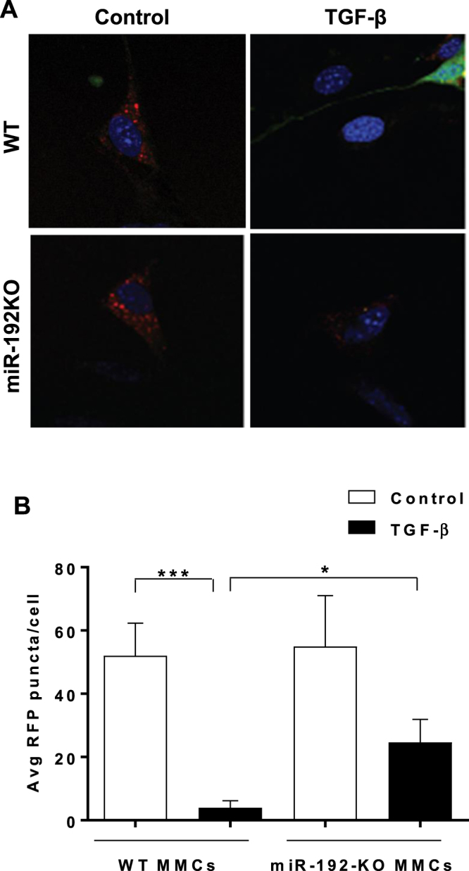
Autophagosome formation is inhibited to a greater extent in WT-MMCs compared to miR-192-KO MMCs following TGF-β treatment. (A) Representative confocal microscopy images showing RFP-LC3 puncta in WT or miR-192-KO MMCs transfected with GFP-RFP-LC3 plasmid (1 µg) followed by TGF-β treatment of these cells for 24 hrs. x1000 (10 × 100 oil) magnification. (B) Bar graph showing quantification of RFP-LC3 puncta in WT and miR-192-KO MMCs. *P < 0.05; ***P < 0.001.