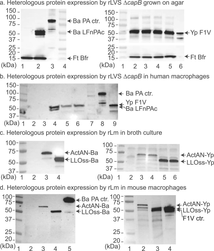
Figure 1.
Expression of heterologous fusion proteins of B. anthracis and Y. pestis by rLVS ΔcapB and rLm ΔactA ΔinlB prfA vaccines grown on agar and in infected macrophage-like cells. (a) Expression of B. anthracis (left) and Y. pestis (right) fusion proteins by rLVS ΔcapB grown on agar. Single colonies of chocolate agar grown rLVS ΔcapB/Ba and rLVS ΔcapB/Yp (4 clones) were lysed in SDS sample buffer and lysates analyzed by Western blotting using a mixture of antibody to B. anthracis PA and to F. tularensis Bfr (left panel) or antibody to Y. pestis LcrV protein followed by antibody to Bfr (right panel). Left panel, lane 1, LVS ΔcapB vector; lane 2, rLVS ΔcapB/Ba; lane 3, PA protein control; lane 4, protein mass standards. Right panel, lane 1, protein mass standards; lanes 2–5, rLVS ΔcapB/Yp; lane 6, monomer of F1-LcrV (F1V) protein control. (b) Expression of fusion proteins by rLVS ΔcapB in infected human macrophage-like cells. Monocytic THP-1 cells seeded on 24-well plates and differentiated in the presence of PMA were left uninfected or infected with LVS ΔcapB, rLVS ΔcapB/Ba or rLVS ΔcapB/Yp; cells were lysed at 24 h post infection, and cell lysates analyzed by Western blotting using a mixture of antibody to B. anthracis PA and to Y. pestis LcrV. Lanes 1 & 7, two different protein standards; lane 2, uninfected control; lane 3, LVS ΔcapB; lane 4, rLVS ΔcapB/Ba; lanes 5 and 6, two clones of rLVS ΔcapB/Yp vaccines; lane 8, B. anthracis PA and degraded proteins; lane 9, Y. pestis F1-LcrV monomer protein and degraded proteins. (c) Expression and secretion of heterologous fusion proteins by rLm vaccines in broth. Culture filtrates of Lm vector or rLm vaccines were analyzed by Western blotting using antibody to B. anthracis PA (left panel) or to Y. pestis LcrV (right panel). Left panel, lane 1, protein mass standards; lane 2, Lm vector; lane 3, rLm/ActAN-Ba; lane 4, rLm/LLOss-Ba. Right panel, lane 1, protein mass standards; lane 2, Lm vector; lanes 3 & 4, two clones of rLm/ActAN-Yp; lanes 5 & 6, two clones of rLm/LLOss-Yp. (d) Expression of heterologous fusion proteins by rLm vaccines in infected mouse macrophage-like cells. Monolayers of J774A.1 cells were not infected or infected with a stationary culture of rLm vaccines similarly as described above in the legend to b . Lysates were subjected to Western blotting analysis using antibody to B. anthracis PA (left) or to Y. pestis LcrV (right). Left panel, lane 1, protein standards; lane 2, uninfected control; lane 3, rLm/ActAN-Ba; lane 4, rLm/LLOss-Ba; lane 5, PA protein. Right panel, lane 1, protein standards; lane 2, rLm/ActAN-Yp; lane 3, rLm/LLOss-Yp; lane 4, F1-LcrV protein control. (a–d) On the left border of each panel are listed the masses of protein standards; on the right border are listed the proteins of interest. Each blot was processed by using the Bio-Rad imaging system (ChemiDoc XRS) and Quantity One software, which allows the overlap of a white-light image, for visualization of the protein standards (a, left panel lane 4 and right panel lane 1; b–d, lane 1), and a chemiluminescent image, for visualization of the antibody-labeled protein bands. The full-length blots in panels b–d are shown in the Supplementary Information (Fig. S1).

