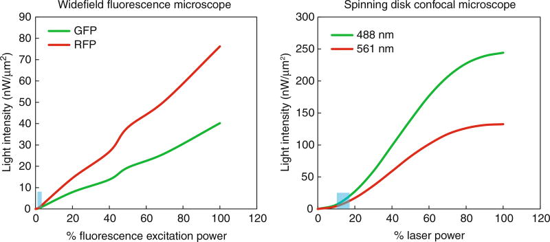
Fig. 1.
Measured light intensity by microscope. Light intensity was measured using an X-cite power meter (Excelitas Technologies) over different settings of transmitted light for the widefield fluorescence microscope and laser power for the spinning disk confocal microscope. The blue boxes represent the range of settings used during live-cell yeast imaging

