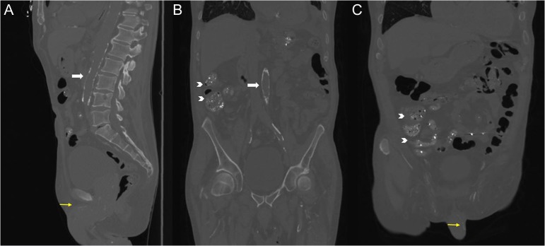
Figure 2:
Sagittal (A), coronal (B) and oblique (C) views of non-contrast CT scan of the abdomen demonstrating extensive calcification of the aorta (white arrows) and pudendal vessels (yellow arrows). The intraluminal densities in the bowel that were incidentally found (indicated by chevrons) are likely due to lanthanum carbonate, which is a metal-based phosphate binder.

