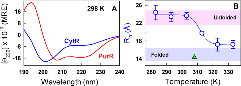
Figure 1.
Disorder and collapse. (A) Far-UV CD spectra of CytR (blue) and PurR (red) at 298 K in mean residue ellipticity units of deg. cm2 dmol−1. (B) Changes in the hydrodynamic radius (Rh) of CytR with temperature extracted from dynamic light scattering measurements. The curves are shown to guide the eye. Shaded regions in pink and blue represent the hydrodynamic radius of disordered and ordered proteins, respectively, from size-scaling expectations (44). The green triangle represents the Rh of CytR from the DNA-bound structure at 308 K calculated using HYDROPRO (45).

