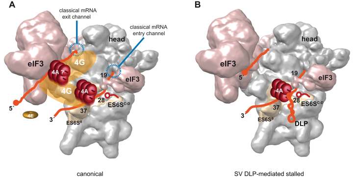
Figure 8.
Model of PIC scanning. (A) Canonical model of PIC scanning with eIF4F complex placed on ES6S region of 40S subunit (EM-5658) (19), interacting with both 3′and 5′regions of mRNA (orange). The scaffolding eIF4G factor is in yellow, interacting with both ES6S (48) and eIF3 as reported before (49,50). In this model two molecules of eIF4A (red rollers) are placed on eIF4G according to previous data (13,15). The 4E subunit of eIF4F (eIF4E) is dissociated from the cap structure upon 48S complex formation as suggested recently (3). (B) SV DLP-mediated stalled PIC showing one eIF4A molecule jammed by DLP structure on ES6S region. The rest of factors and details were omitted for simplicity. The position of VIColigo-4 is also shown (red).

