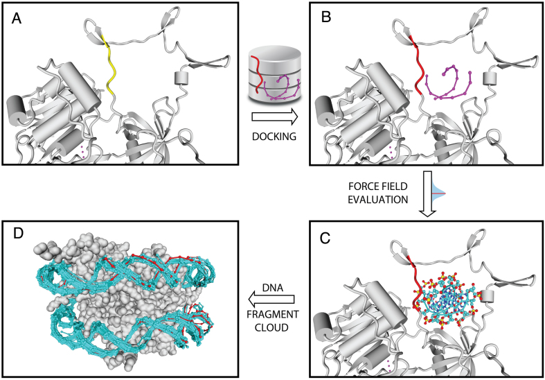
Figure 2.
Docking procedure: (A) a protein fragment (yellow) is used to query the pepX database for a compatible fragment; (B) the retrieved pepX fragment (red) is superimposed on the yellow one placing the associated DNA fragment (dnaX, purple); (C) backbone dnaX atoms are evaluated with the PADA1 force field; (D) an example with a histone octamer showing all dnaX docked models (cyan) fully covering the crystallographic DNA (red).

