Figure 6.
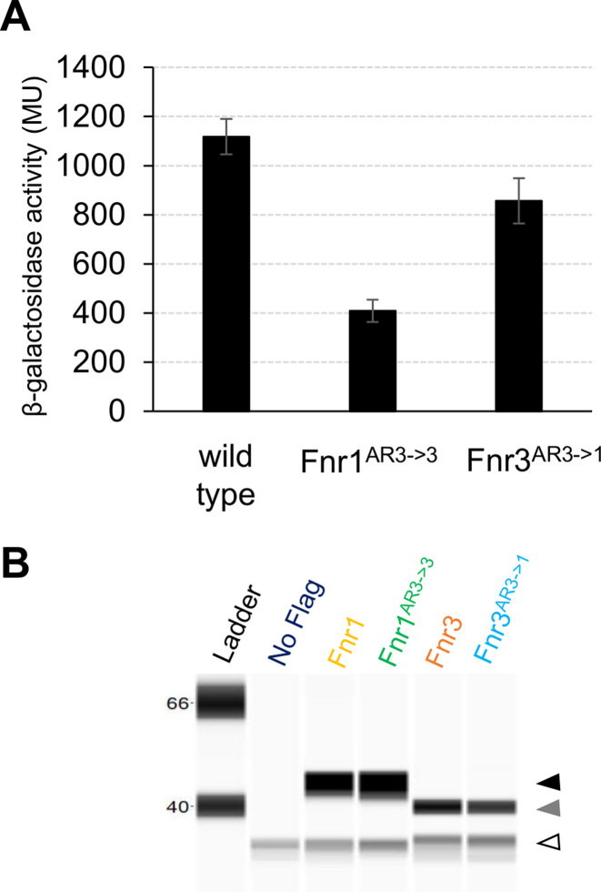
Role of Activating Region 3 in transcriptional activation by Fnr proteins. (A) Activity of the native pfnr1::lacZ fusion in the wild type strain of H.seropedicae in comparison with strains carrying specific activating region domain swaps. The swapped domain strains are Fnr1AR3→3 (Fnr1 with Fnr3-like activation region 3) and Fnr3AR3→1 (Fnr3 with Fnr1-like activation region 3). β-galactosidase activity, in Miller Units, was assayed as described in Materials and Methods using cultures incubated for eight hours after the switch from high to low oxygen. The error bars represent the standard error for three independent biological replicates. (B) Levels of the swapped domain proteins are similar to the cognate wild type Fnr protein. 3xFlag tagged versions of the Fnr1 (yellow), Fnr1AR3→1 (green), Fnr3 (orange), and Fnr3AR3→1 (blue) proteins were used to verify the protein levels using the Simple Wes system as described in Materials and Methods. The black arrowhead indicates Fnr1, the grey arrowhead indicates Fnr3, and the white arrowhead represents the cross reacting protein from H. seropedicae. The protein extracts were prepared from the same cultures used for the β-Galactosidase activity measurements shown in A.

