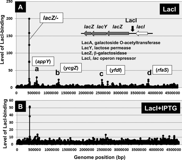
Figure 2.
Genomic SELEX pattern of LacI. The Genomic SELEX screening was performed for LacI in the absence (A) and presence (B) of the inducer IPTG. In the absence of IPTG, a single peak was detected within the spacer between the lacZYA and lacI operons. The appearance of low-level peaks was not reproducible, suggesting no specific binding to these sites. By adding increased concentrations of IPTG, the level of LacI binding to the single target decreased gradually. Minor peaks in (A) are located inside ORF of appY (a), ycgZ (b), yfdI (c) and rfaS (d). These one-point peaks might be non-specific background noise because ∼300 bp long SELEX fragments should bind to two or more consecutive 60 bp long probes aligned at 105 bp intervals on the tilling array used (25).

