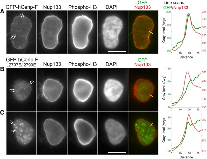
Figure 4. L2797E/I2799E mutations prevent GFP‐hCenp‐F targeting to the NE in late G2/prophase cells.

-
A–CHeLa‐E cells transiently transfected with GFP‐hCenp‐F‐WT (A) or GFP‐hCenp‐F‐L2797E/I2799E (B, C) were fixed 1 day after transfection and immunolabeled with the indicated antibodies. A single plane from widefield acquisitions of prophase cells (identified based on phospho‐histone H3 staining and persistence of a NE) is shown. Scale bars, 10 μm. Arrows point to kinetochores labeled by GFP‐hCenp‐F. Line scans (yellow lines on images, plotted from the cytoplasm toward the nucleoplasm, distances in pixels) measuring the intensity of GFP‐hCenp‐F (green lines) and Nup133 (red lines) signals in late G2/prophase cells reveal the peak of GFP‐hCenp‐F‐WT that co‐localizes with Nup133 at the NE (A). In contrast, either no enrichment (B) or a very minor accumulation (C) is detectable in GFP‐hCenp‐F‐L2797E/I2799E‐transfected cells.
