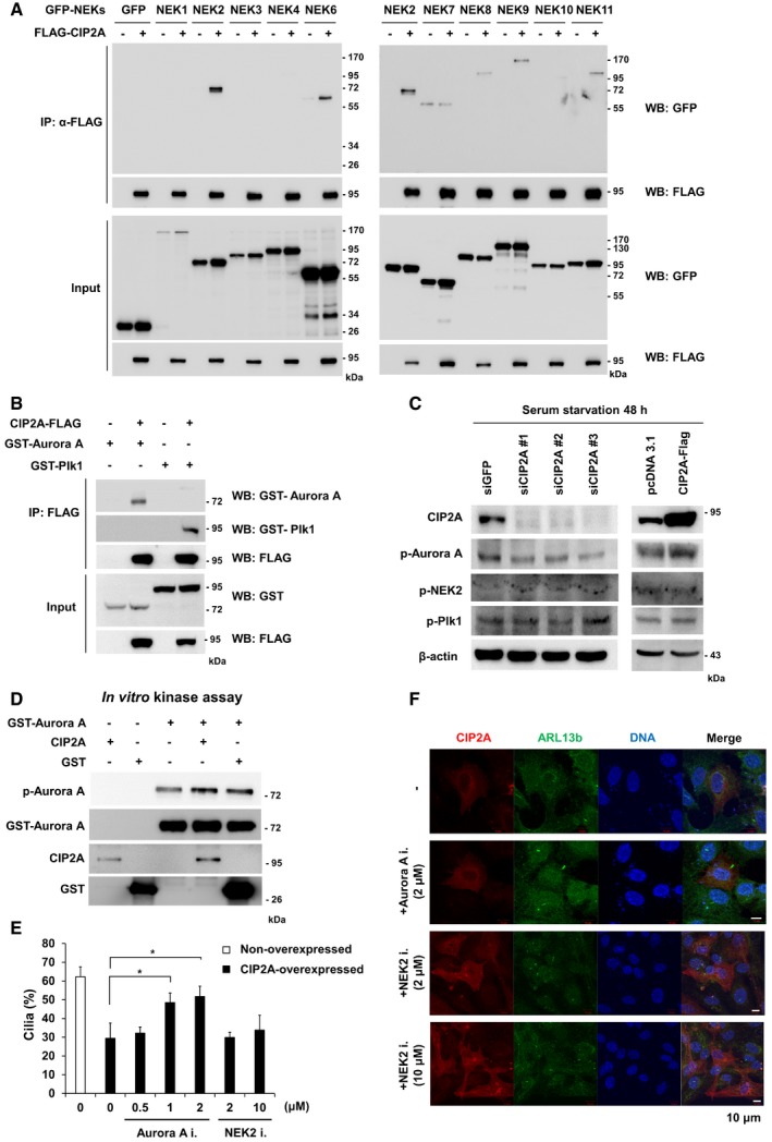
Figure 3. CIP2A interacts with and activates Aurora A, which disassembles primary cilia.

- HEK293 cells were used for a co‐immunoprecipitation analysis between FLAG‐tagged CIP2A and GFP‐tagged NEK family proteins. HEK293 cells lysates were immunoprecipitated with anti‐FLAG antibodies, and IP proteins were analyzed by immunoblot.
- FLAG‐tagged CIP2A constructs were coexpressed with the GST‐tagged Plk1 construct or the GST‐tagged Aurora A construct in HEK293 cells. FLAG‐tagged proteins were immunoprecipitated with anti‐FLAG antibodies, and the co‐immunoprecipitation of GST‐tagged proteins was determined by immunoblot.
- RPE1 cell lysates after siRNA transfection and serum starvation for 48 h were used for immunoblot analyses.
- Aurora A activity was measured using an in vitro kinase assay with recombinant proteins. After the kinase reaction, samples were subjected to SDS–PAGE. Samples were transferred onto a nitrocellulose membrane and processed for immunoblot using phosphorylation‐specific Aurora A antibodies.
- The percentage of CIP2A‐overexpressed and non‐overexpressed cells with primary cilia was evaluated after treatment with the Aurora A inhibitor MLN 8237, or the NEK2 inhibitor Rac‐CCT 250863 (n > 14 cells/condition) under serum‐starved condition. The average of three independent experiments is shown, with error bars representing s.d. *P < 0.05 compared with MLN 8237 non‐treated cells (one‐tailed Student's t‐test).
- Immunofluorescence images of RPE1 cells transfected with CIP2A‐FLAG following serum starvation for 48 h and treatment with MLN 8237 or Rac‐CCT 250863 for 24 h. Primary cilia were stained with antibodies specific for ARL13b (red). DNA was stained with DAPI (blue). Shown are the maximum projections from z stacks of representative non‐treated or treated transfected cells. Scale bar = 10 μm.
Source data are available online for this figure.
