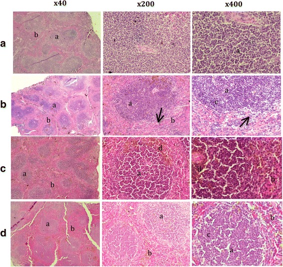
Fig. 2.

Splenic section obtained after administration in mice from the following groups: a normal, b non-treatment, c combination of antibiotic and root extract, d C. mongolica root extract only. a - white pulp, b - red pulp, c - lymphocytes in different stages of development, d - haemosiderin pigmentation, ↗ - lymphocyte necrosis. Haematoxylin and eosin stain, magnification 400× and 40×
