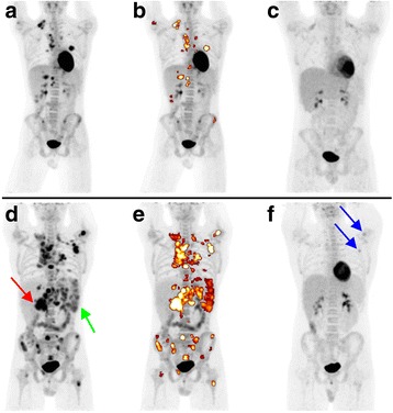
Fig. 1.

Patient examples of low and high MTV. Representative examples of FDG-PET maximum intensity projections (MIP) of two patients with stage IV disease before induction therapy (a, b + d, e) and at ERA (c, f). In the middle column (b, e), the delineated pretherapeutic MTV is colored (high activity: white, low activity: brownish). a-c: A 17-year-old male with stage IV disease (liver, lung) and AR who had a low MTV (51 ml). d-f: A 17-year-old male with stage IV disease (skeletal) and IR who showed a high MTV (792 ml); please also note the large lymph node mass at the liver hilus (red arrow) and extensive splenic involvement (green arrow). At ERA, considerable FDG uptake (Deauville score 4) can still be detected especially in a left axillary lymph node and the left humerus (blue arrows)
