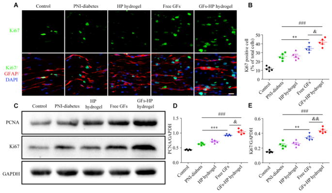
Fig. 7.
GFs-HP hydrogel facilitates SCs proliferation. A. The double immunofluorescence staining of Ki67 (green) with the GFAP (red) to label proliferating SCs in longitudinal sciatic nerve sections, Scale bar=50μm; B. The percentages of cells double-positive for Ki67 and GFAP out of total DAPI positive cells (representing proliferation rate of SCs); C. The protein levels of Ki67 and PCNA at 30 days after injury by western blotting; D, E. Densitometric analyses of PCNA and Ki67, respectively. GAPDH was used for band density normalization. Data presented as mean±SEM, n=5 for each group. Free GFs vs PNI-diabetes: **P <0.01, ***P <0.001, GFs-HP hydrogel vs PNI-diabetes: ###P<0.001, GFs-HP hydrogel vs Free GFs: &P <0.05, &&P <0.01. (For interpretation of the references to color in this figure legend, the reader is referred to the Web version of this article.)

