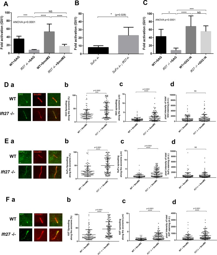
FIGURE 1:
IFT27 is fully integrated in hedgehog signaling pathway. (A) qRT–PCR quantification of Gli1 expression in wild-type and Ift27 knockout cells activated by SAG or by the transfection of SmoM2. Five independent analyses were performed. (B) qRT–PCR quantification of Gli1 expression in SuFu knockout cells or SuFu, Ift27 double knockout cells. Five independent analyses were performed. (C) qRT–PCR quantification of Gli1 expression in wild-type and Ift27 knockout cells activated by SAG or by the transfection of constitutively active form of Gli2 (Gli2ΔN). Five independent analyses were performed. (D–F) Localization of hedgehog effectors in wild-type and Ift27 knockout cells. Immunofluorescence staining of Gli2 (Da), SuFu (Ea), and Kif7 (Fa). Quantification of the spreading pattern presented as percentage of the axoneme covered by the staining (Db, Eb, Fb) or length (Dc, Ec, Fc). Quantification of total fluorescence intensity for each of the cilia (Dd, Ed, Fd). In each case, ∼120 cilia were measured. Data were analyzed using one-way Kruskal–Wallis test (analysis of variance [ANOVA]) with Tukey’s multiple comparison test (A, C), two-tailed Kolmogorov–Smirnov test (B), or Mann–Whitney test (C, Db, Dc, Dd, Eb, Ec, Ed, Fb, Fc, Fd). Error bars are SD. Bars, 1 micron.

