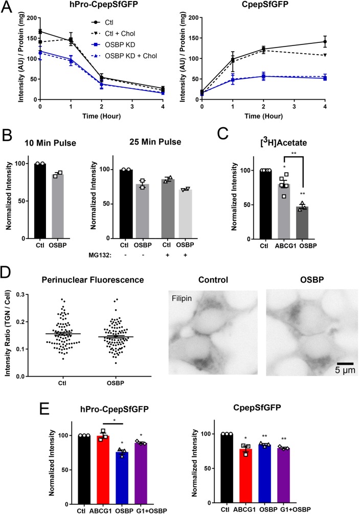
FIGURE 7:
OSBP depletion reduces synthesis of hPro-CpepSfGFP and decreases CpepSfGFP accumulation but does not amplify the effects of ABCG1 knockdown. (A) Effect of OSBP knockdown and pretreatment with exogenous cholesterol on biosynthetically labeled hPro-CpepSfGFP and CpepSfGFP. hPro-CpepSfGFP levels in OSBP KD (+/− cholesterol) are significantly different from control (+/− cholesterol) at 0 and 1 h. CpepSfGFP levels (+/− cholesterol) are significantly different in OSBP KD from control (+/− cholesterol) at 1 h and later. Significance determined by two-way ANOVA; n = 3. (B) Neither shortening the labeling period (left) nor including the proteasomal inhibitor MG132 (right) affects the level of accumulation of hPro-CpepSfGFP. Data (intensity/mg protein) are normalized to the control (n = 2, each in duplicate). (C) Cholesterol biosynthesis as measured by [3H]acetate incorporation showing a slight decrease in ABCG1-depleted cells and strong decrease in OSBP-depleted cells. Quantification from scintillation counting of [3H]cholesterol separated by thin-layer chromatography; n = 3–5. (D) Modest but not significant decrease in filipin fluorescence concentrated perinuclearly in OSBP KD as compared with control. Fixed and stained cells are shown in reverse contrast to highlight perinuclear fluorescence, which was quantified and normalized to total cell fluorescence; control cells, # = 84; OSBP KD cells, # = 91 cells. Significance determined by Student’s t test. (E) Combined knockdown of ABCG1 and OSBP does not increase the loss of CpepSfGFP beyond the level observed with either knockdown alone. Quantification from Western blots; n = 3. Data are presented as mean ± SEM; p values are determined by Student’s t test; *, p < 0.05; **, p < 0.01.

