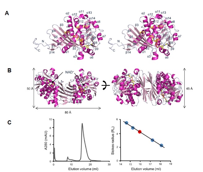
Fig. 1. Structure of Streptomyces pristinaespiralis LCD in complex with NAD+.

(A) Stereo ribbon diagram of S. pristinaespiralis LCD (SpLCD)/NAD+ monomer. Helices are light pink, strands are magenta, and loops are grey. The yellow stick represents NAD+. All figures, including protein structures, were drawn using PyMOL software (The PyMOL Molecular Graphics System, http://www.pymol.org). (B) Ribbon diagram of SpLCD/NAD+ dimer. The height of the structure was measured after rotating the structure 90 degrees. (C) Analyti-calgel filtration profile of SpLCD. Standard curve generated using molecular weight markers. The positions of the molecular weight markers for β-amylase, alcohol dehydrogenase, carbonic anhydrase, and cytochrome C are indicated with blue dots. The position of SpLCD is marked with a red dot.
