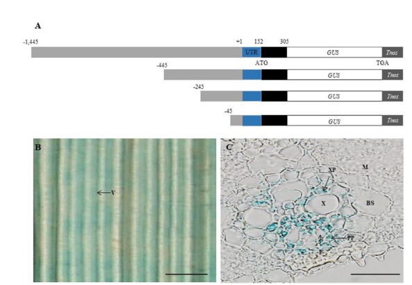
Fig. 1. Schematic representation of Hd3a promoter-GUS reporter constructs and histochemical localization of GUS.

(A) Hd3a genomic fragments comprising −1,445, −445, −245, and −45 bp promoter regions (grey box), the 152-bp 5′ UTR (blue box) and 153-bp coding region of Hd3a (black box) were connected to GUS coding region and nopaline synthase terminator (Tnos). (B) Leaf blade of −1,445 Hd3a promoter–GUS transgenic at ZT 1 on 35 DAG. (C) Transverse section of leaf blade in Panel B. BS, bundle sheath cells; M, mesophyll cells; P, phloem; PP, phloem parenchyma; V, vascular bundle; X, xylem; XP, xylem parenchyma. Scale bars =1 mm (B) and 20 μm (C).
