
Fig. 4. Histochemical localization of GUS activity in roots at five developmental stages.
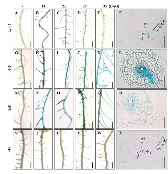
(A–E) Roots of transgenic plants carrying −1,445 Hd3a promoter–GUS at 7 (A), 14 (B), 21 (C), 28 (D), and 35 (E) DAG. (G–K) Roots of transgenic plants carrying −445 Hd3a promoter–GUS at 7 (G), 14 (H), 21 (I), 28 (J), and 35 (K) DAG. (M–Q) Roots of transgenic plants carrying −245 Hd3a promoter–GUS at 7 (M), 14 (N), 21 (O), 28 (P), and 35 (Q) DAG. (S–W) Roots of transgenic plants carrying −45 Hd3a promoter–GUS at 7 (S), 14 (T), 21 (U), 28 (V), and 35 (W) DAG. Cross-sections of roots at 35 DAG from −1,445 Hd3a promoter–GUS (F), −445 Hd3a promoter–GUS (L), −245 Hd3a promoter–GUS (R), and −45 Hd3a promoter–GUS (X). Co, cortex; CX; central xylem; En, endodermis, Ep, epidermis; Ex, exodermis; P, phloem; Sc, sclerenchyma; V, vascular bundle; X, xylem. Scale bars = 50 μm in Panels F, L, R, and X; 5 mm in others.