
Figure 1.
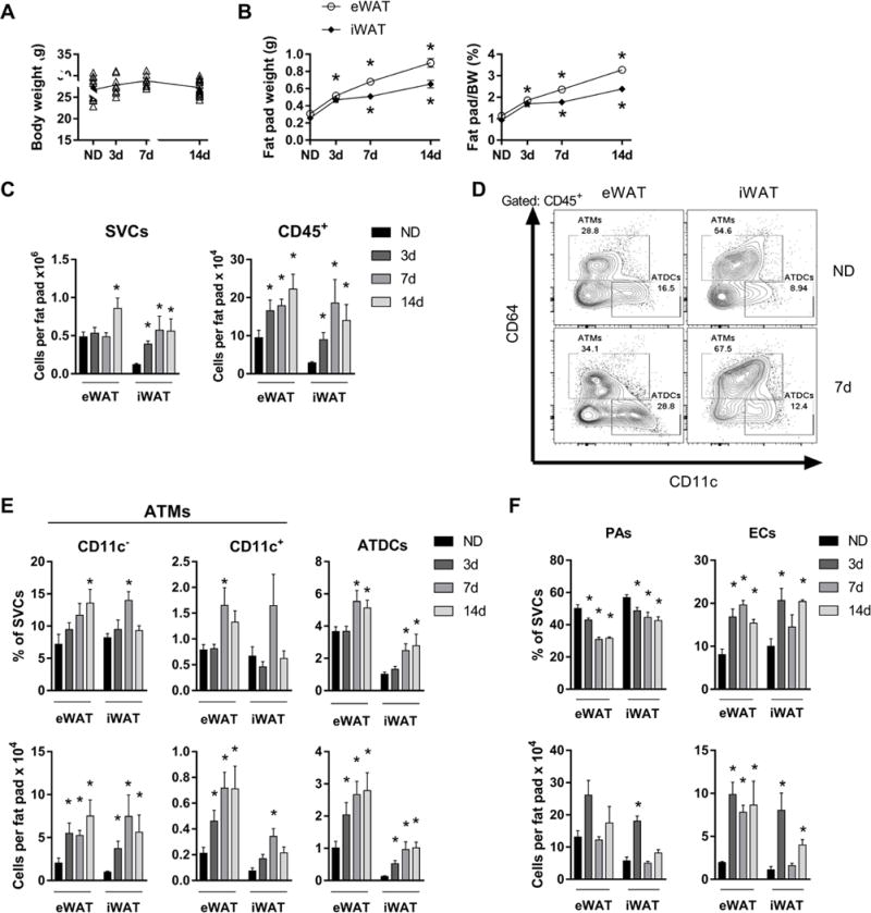
Increased ATMs, ATDCs, and endothelial cells with rapid WAT expansion. Body weights (A) and fat pad weights normalized to body weight (BW) (B) of mice fed ND or 60% HFD for 3-14 days. (C) Quantity of SVCs and CD45+ leukocytes in eWAT and iWAT. (D) Representative flow plots showing ATM and ATDC frequencies in eWAT and iWAT for ND and mice fed HFD for 7 days. (E and F) Frequency and quantity of CD11c− ATMs, CD11c+ ATMs, ATDCs, preadipocytes (PAs), and endothelial cells (ECs) in ND and in mice fed HFD for 3-14 days. *, p<0.05.