Figure 3.

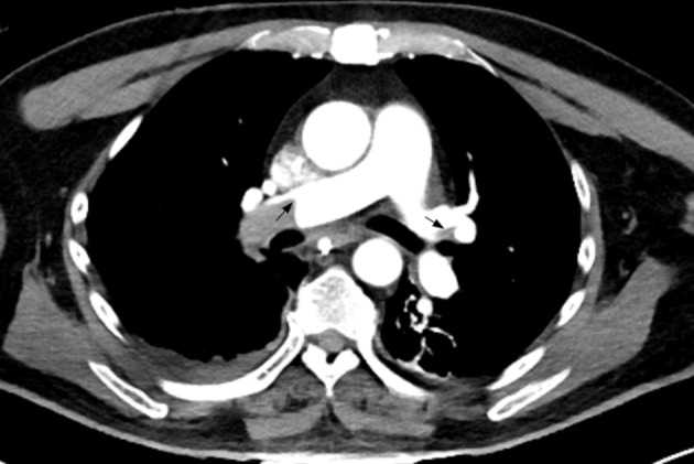
Coronal section of the chest CT angiography showing bilateral pulmonary arterial filling defects (large right main stem and small left segmental, marked by arrow) suggestive of pulmonary embolism.

Coronal section of the chest CT angiography showing bilateral pulmonary arterial filling defects (large right main stem and small left segmental, marked by arrow) suggestive of pulmonary embolism.