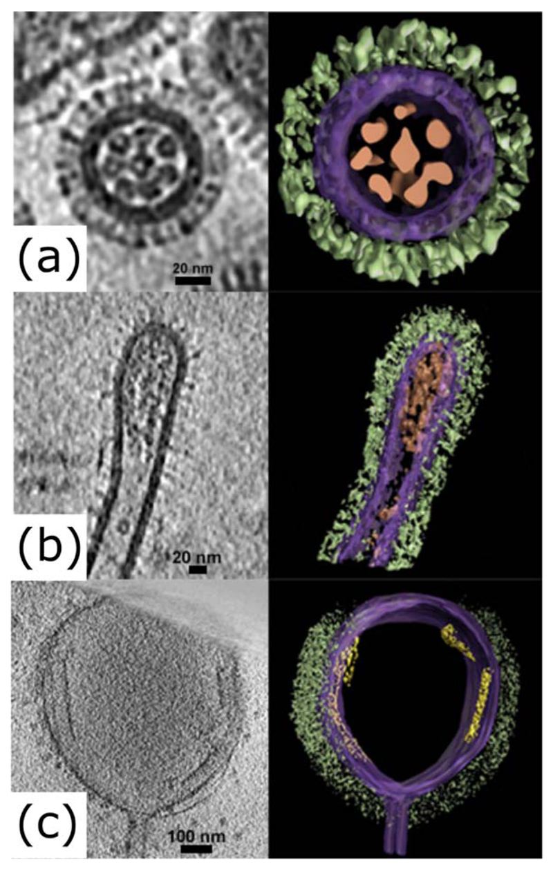
Figure 2. Bacilliform and filamentous influenza virions at high resolution.
Electron tomograms of influenza virions, showing slices (left panels) and segmented images (right panels) of (a) a transverse section of a bacilliform virion, (b) a longitudinal section of the tip of a filamentous virion and (c) a longitudinal section of an Archetti body at the end of a filamentous virion. Images were manually segmented and coloured to show viral glycoproteins (green), membrane and associated matrix (purple), genome (brown) and putative free M1 sheets (yellow). Tomograms were obtained as part of a previous study (Vijayakrishnan et al., 2013) and manually segmented using Amira (TGS).

