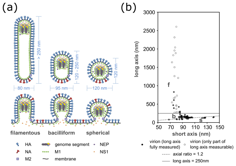
Figure 3. Dimensions of influenza virions.
The dimensions of influenza virions, shown (a) as a schematic of budding and released virions, with typical sizes indicated, and (b) as measurements of purified influenza A/Udorn/72 virions. For (a) it should be noted that the incorporation of NS1 and NEP has so far only been examined in spherical virions, and their general incorporation is inferred from this. For (b) measurements of 96 virions were taken by cryoelectron microscopy (data replotted from Vijayakrishnan et al., 2013). Open circles indicate filaments which extended beyond the field of view and so are longer than measured. Spherical virions (s) are distinguished from bacilliform virions (b) by having an axial ratio less than 1.2 (dashed line); filaments (f) are distinguished from bacilliform virions by having a length greater than 250 nm (solid line).

