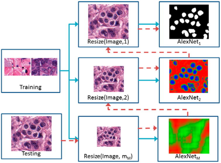
Figure 1.

High-level flow chart illustrating RADHicaL. Training images (blue paths) are resized to train M different AlexNet networks at each of the different resolutions m. Test images (red path) are initially reduced to the smallest magnification, where a threshold is applied to the classifier's probability mask so that only relevant pixels are mapped into the next highest resolution for further analysis.
