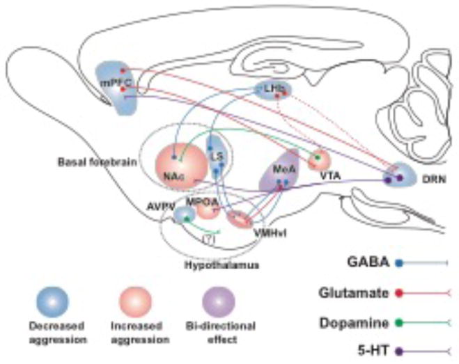
Figure 1. Neural Circuitry of Aggression.
Schematic map of the brain areas and their neuronal connection that are involved in aggressive behavior in mice reviewed in this paper. Areas that stimulate aggression are shaded in “pink” and areas that suppress aggression are shaded in “blue”. Medial amygdala (MeA) is shaded in “violet” and since it can both stimulate and suppress aggression depending on neuronal output that is optogenetically stimulated. AVPV: anteroventral periventricular nucleus of hypothalamus. DRN: dorsal raphe nucleus. LHb: lateral habenula. MeA: Medial amygdala. MPOA: medial preoptic area. NAc: nucleus accumbens. VMHvl: ventrolateral area of ventromedial hypothalamus. VTA: ventral tegmental area. (Dashed line denotes neuronal pathways that have been identified but not studied in the context of aggression or aggression-seeking behavior. The question mark [?] reflects the fact that although highly possible, dopaminergic nature of these neurons yet to be proven).

