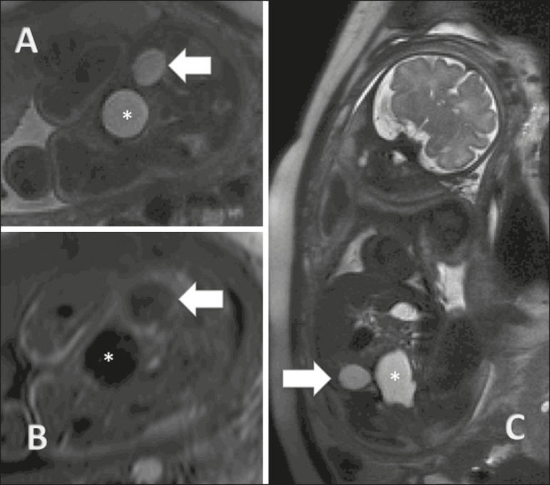
Figure 4.
Ovarian cyst in a fetus at 32 weeks of gestation. A: Axial T2-weighted sequence of the fetal pelvis showing a hyperintense signal in the right adnexal region. B: Axial T1-weighted sequence of the fetal pelvis, showing a well-defined area of signal hypointensity, with homogeneous contours, in the right adnexal region. C: Coronal T2-weighted sequence showing a cyst with a hyperintense signal in the right adnexal region. Ovarian cyst (arrows) and fetal bladder (asterisks).

