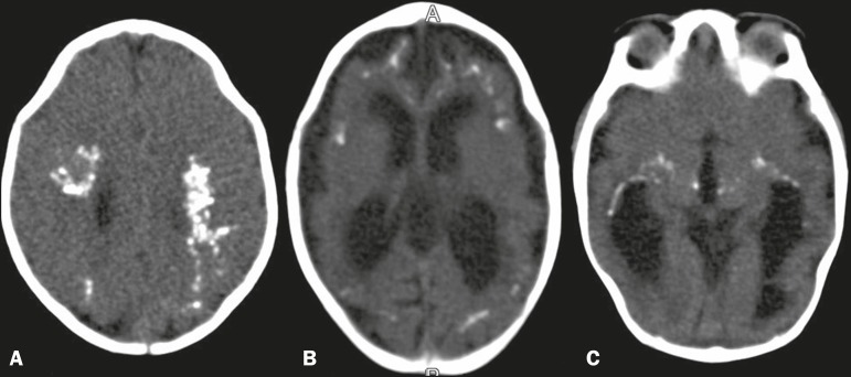
Figure 1.
CT scan showing parenchymal calcifications in several locations in different patients. A: In the deep white matter and in cortical-subcortical regions, at the level of the corona radiata, assuming a confluent aspect. B: In cortical-subcortical regions, at the level of the lateral ventricles. C: Calcifications in the thalamus and in the capsulonuclear regions. Note the narrowing of the frontal bones, in A, and the small left frontal subcortical cyst, in B.

