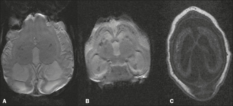
Figure 2.
MRI. A,B: Volumetric gradient-echo sequences showing foci of signal hypointensity in the cortical-subcortical regions, thalamic, and capsulonuclear regions. C: Volumetric fast spoiled gradient-echo T1-weighted sequence, of the same patient depicted in B, showing foci of signal hyperintensity, confluent in the cortical-subcortical regions, corresponding to the foci of signal hypointensity seen in the gradient-recalled echo sequence.

