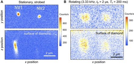
Fig. 2. Strobed confocal microscopy of NV centers.

(A and B) NV1 and NV2 when stationary (A) and when rotating at high speed (B). Upper panels are x-y plane scans, whereas lower panels are x-z plane scans into the diamond. The color bar describes count rate at the experimental duty cycle of D = 0.67% (peak counts of 1 × 10⁵ recorded for D = 1). The stationary confocal images of NV1 and NV2 are strobed synchronously with an external 3.33-kHz signal from a function generator to yield an equivalent duty cycle to the rotating case. Counts are integrated for 200 ms at each pixel with the laser pulse duration tpulse = 2 μs. (B) The optical illumination and collection are pulsed to be synchronous with the 3.33-kHz rotation of the diamond; blurring resulting from period jitter and wobble of the rotation is evident. NV1 was used for the remainder of the measurements described in this work.
