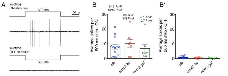
Figure 7. Electrophysiological recordings from wildtype and emx2 mutant afferent neurons.
(A) Example of wildtype spikes during a 500 ms ON and OFF waterjet stimulus. (B) Quantification of the average spikes per 10, 500 ms step stimuli in the direction of sensitivity (ON-stimulus). For wildtype, the A > P and P > A afferent neuron spike numbers were pooled. A similar number of spikes were detected in wildtype, emx2 ko and emx2 gof neurons. (B’) Quantification of the average spikes for the same neurons as in (B), except in the opposite direction (OFF-stimulus). The number of afferents recorded: WT, n = 15; emx2 ko, n = 8; emx2 gof n = 7, obtained from a minimum of three independent experiments. The one-way ANOVA was used for the comparison in (B) and (B’).

