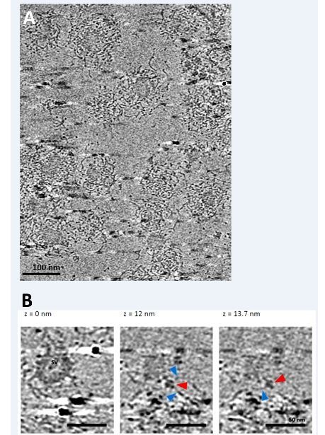
Author response image 2. Cryo-electron tomography on partially uncoated CCVs reveals that clathrin coat-vATPase interactions exist.

(A) Electron micrographs of partially uncoated CCVs (in tightly packed CCVs, vATPase could not be detected). (B) Zoomed image of two vesicles at different views, as indicated. Red arrowhead points presumably to vATPase, blue arrowheads to clathrin filaments. See cryo-electron tomography Video for a better view.
