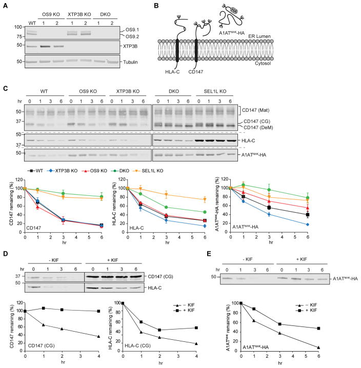
Figure 1. Complex roles of OS9 and XTP3B in glycoprotein ERAD.
(A) Immunoblot analysis of OS9 and XTP3B in cell lysates from wild type (WT) and two independent clonal knockout lines for each genotype. (B) Schematic representation of the topology and glycosylation status of the glycoproteins examined in this study. (C) Turnover of endogenous and exogenous ERAD substrates. Untransfected (CD147 and HLA-C) or transfected (A1ATNHK-HA) cells were treated with the protein synthesis inhibitors cycloheximide or emetine (PSI-chase) for the indicated times, and glycoprotein levels were determined by immunoblotting with the indicated antibodies. Mat=mature; CG=core-glycosylated; DeM=demannosylated. Quantification represents mean±SEM (n=3). (D and E) Effect of kifunensine on glycoprotein turnover. Untransfected (D) and A1ATNHK-HA transfected (E) cells were treated (~16 hrs) with kifunensine (KIF) prior to PSI-chase and immunoblot with the indicated antibodies. The vertical line indicates where a lane with protein marker was cut for simplicity and independent immunoblots are indicated by the dashed horizontal lines.. See also Fig. S1.

