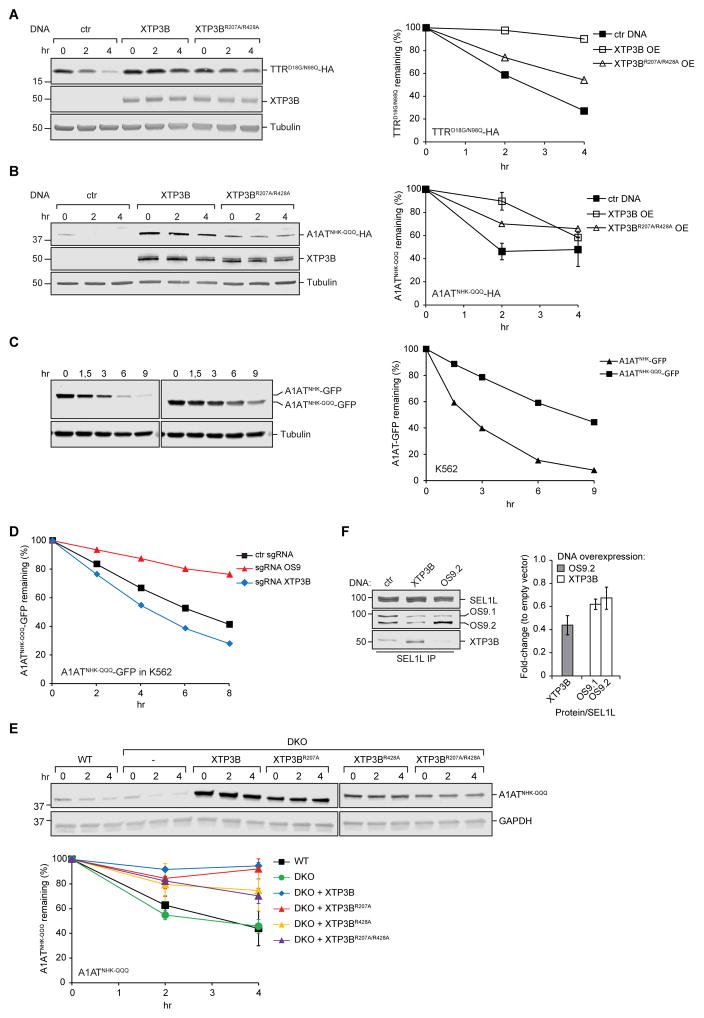
Figure 5. Inhibition of non-glycosylated protein ERAD by XTP3B MRH2.
(A, B) PSI-chase analysis 48 hrs after transfection with TTRD18G/N98Q-HA (A), A1ATNHK-QQQ-HA (B), XTP3B, XTP3BR207A/R428A or empty vector in wildtype cells. Quantification is based on one (A) or two (B) independent experiments for the empty vector and wild type XTP3B and one experiment for XTP3BR207A/R428A (B). Error bars represent STDEV. (C) Expression of A1ATNHK-GFP or A1ATNHK-QQQ-GFP in K562 cells was induced by growing cells in the presence of 0.1 μg/mL doxycycline (dox) for 16 hrs. Cells were subsequently treated with emetine for the indicated times. Protein turnover was determined by immunoblotting with an anti-GFP antibody. n=1. (D) K562 cells stably expressing dox-inducible A1ATNHK-QQQ-GFP and the indicated sgRNA were treated with 0.1 μg/mL doxycycline (dox) for 16 hours. Cells were subsequently treated with emetine and GFP median fluorescence intensity was measured by flow cytometry at the indicated times. Quantification is based on one experiment. (E) PSI-chase analysis of A1ATNHK-QQQ-HA in WT, DKO, and DKO cells stably expressing the indicated XTP3B constructs. Quantification represents the mean±STDEV (n=2). (F) Competition between OS9 and XTP3B for binding to SEL1L. Endogenous SEL1L was immunoprecipitated from 1% Triton X-100 lysates of wild type HEK293 cells transfected with OS9.2, XTP3B, or empty vector. SEL1L complexes were analyzed by immunoblotting with the indicated antibodies. Quantification of XTP3B and OS9 normalized to SEL1L is represented as mean±SEM (n=3). See also Fig. S4.

