
Figure 3. SMG derived Sox9-lineage labeled cells repair SE and contribute to both luminal and basal cells following injury.
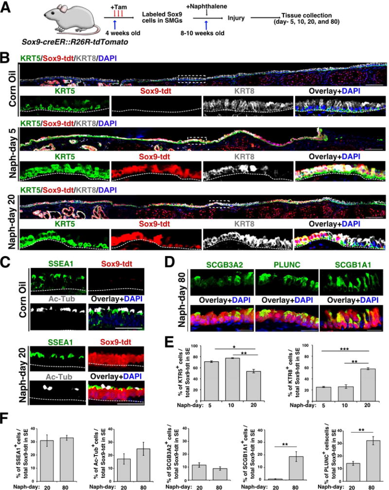
(A) Schematic of Sox9-tdt lineage trace, naphthalene injury and tissue collection.
(B) Co-staining of KRT5 (green) and KRT8 (grey) on tracheal sections collected from control and injured mice (day-5 and 20). Scale bar: 50μm. Dotted box indicates region shown at high magnification in panels below. The dotted line separates SE and mesenchyme. Scale bar: 50μm.
(C) Tracheal sections from Sox9-tdt naphthalene injured (day-20) or control mice stained for SSEA1 – secretory cell marker (green), AcTub (acetylated tubulin) – ciliated cell marker. Sox9-tdt (red) indicates Sox9-lineage labeled cells, nuclear DAPI (blue). Scale bar: 50μm.
(D) Staining of secretory cell markers: PLUNC, SCGB1A1 and SCGB3A2 (green) on day-80 naphthalene injured Sox9-tdt (red) mice. Nuclear DAPI (blue). Scale bar: 20μm.
(E) Quantification of KRT5+ and KRT8+ cells among total Sox9-tdt+ cells in SE on day-5, 10, and 20 post naphthalene injury. Data are shown as mean ± SEM (n=3; *p ≤ 0.05; ** p ≤ 0.01; ***p ≤ 0.001).
(F) Quantification of SSEA1+, Ac-Tub+, SCGB3A2+, SCGB1A1+, PLUNC+ cells among total Sox9-tdt+ cells in SE on day-20, and 80 post naphthalene injury. Data are shown as mean ± SEM (day 20: n=3, day 80: n=4 for all markers except SCGB1A1 (n=3); *p ≤0.05; **p ≤ 0.01; ***p ≤ 0.001).
See also Figure S3.