
Figure 5. MEC-derived SE basal-like cells proliferate, engraft and generate basal and luminal cell types in ex vivo engraftment models.
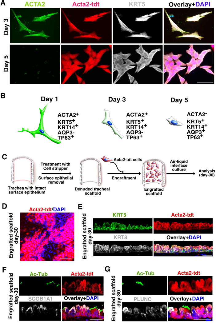
(A) Myoepithelial cells of SMG display SE basal cell characteristics in culture. Cultured cells on day-3 and day-5 co-stained with ACTA2 (green) and KRT5 (grey). Acta2-tdt (red) and nuclear DAPI (blue). Scale bar: 50μm.
(B) Schematic depicting the gradual loss of ACTA2 expression in SMG-derived MECs in culture.
(C) Schematic representation of engraftment of cultured MECs on a denuded tracheal scaffold. Isolated, cultured myoepithelial cells were seeded on denuded trachea followed by air-liquid interface culture and analysis.
(D) Whole mount IHC analysis for tdt-expressing engrafted cells on tracheal scaffold. Scale bar: 50μm.
(E) Co-staining of KRT5 (green) and KRT8 (grey) on trachea engraftment sections. Acta2-tdt (red) and nuclear DAPI (blue). n=3. Scale bar: 20μm
(F) IHC for Ac-Tub (green) and SCGB1A1 (grey) on tissue sections from Acta2-tdt (red) expressing cells engrafted on tracheal scaffold. Nuclear DAPI (blue). Scale bar: 20μm
(G) Sections from engrafted scaffolds expressing Acta2-tdt (red) co-stained with Ac-Tub (green) and PLUNC (grey). Nuclear DAPI (blue). Scale bar: 20μm
See also Figure S5.