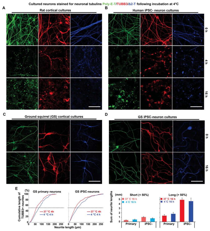
Figure 1. Species-dependent Differences in Neuronal Microtubule Cold Stability.
(A) Immunofluorescence of TUBB3 (red), polyglutamylated tubulin (poly-E-T; green), or delta2-tubulin (Δ2-T; blue) in cultured rat primary cortical neurons incubated at 4°C for various durations. Scale bar: 40 μm.
(B) Immunofluorescence of TUBB3, poly-E-T, or Δ2-T in cultured human induced pluripotent stem cell-derived neurons (iPSC-neurons) incubated at 4°C for various durations. Scale bar: 40 μm.
(C) Immunofluorescence of TUBB3, poly-E-T, or Δ2-T in cultured 13-lined ground squirrel (GS) primary cortical neurons incubated at 4°C for various durations. Scale bar: 40 μm.
(D) Immunofluorescence of TUBB3, poly-E-T, or Δ2-T in GS iPSC-neurons incubated at 4°C for various durations. Scale bar: 40 μm. For other cell types differentiated from GS iPSCs, see also Figure S1.
(E) Cumulative plots and quantification of the lengths of manually traced TUBB3-positive microtubules (see STAR METHODS) from GS cortical primary neurons (n = 5 experiments each at 37°C and 4°C; cultures derived from 4 GS pups) and GS iPSC-neurons (n = 6 experiments each at 37°C and 4°C; cultures derived from 2 GS iPSC lines). Cumulative lengths of short (neurite lengths < 50% of the cumulative plots) and long microtubules (neurite lengths > 50% of the cumulative plots) were quantified separately. No significant difference in microtubule length attributed to cold temperature was found (p > 0.05; Student’s t-test for two-group comparison).

